
About
Service available to small and large animal patients.
The Medical Imaging Service at the WCVM Veterinary Medical Centre provides expertise and technologies for patients in the Small Animal Clinic as well as in the Large Animal Clinic.
WCVM’s team of medical imaging specialists and support staff have experience with a variety of specialized equipment — expertise that enhances the management of cases at the veterinary medical centre.
The WCVM's Medical Imaging Service offers the following services:
- digital radiography
- ultrasonography
- magnetic resonance imaging (MRI)
- computed tomography (CT)
- positron emission tomography-computed tomography (PET-CT)
- nuclear scintigraphy
- imaging of canine hips and elbows for certification with Orthopedic Foundation for Animal (OFA)
- pregnancy examinations
The WCVM Field Service team is also equipped with portable ultrasound and digital radiograph units for pregnancy examinations, lameness case and on-farm diagnostics.
Magnetic Resonance Imaging (MRI)
Magnetic resonance imaging (MRI) is an excellent tool for imaging soft tissues. This noninvasive imaging technology differentiates between various types of soft tissues — not just soft tissue and bone.
It also allows medical imaging specialists to determine whether soft tissue is normal or if it’s something that has abnormal characteristics such as inflammatory tissue or cancer.
The WCVM Veterinary Medical Centre has two MRI units — one for small animal patients and a standing MRI unit for equine patients.
Small Animal MRI
Medical imaging specialists use the small animal MRI unit to:
- diagnose spinal and brain diseases as well as other conditions of soft tissues
- diagnose musculoskeletal diseases (such as muscle and tendon injuries)
- determine the extent of cancer and plan the patient's treatment (surgery, radiation or chemotherapy)
Standing MRI
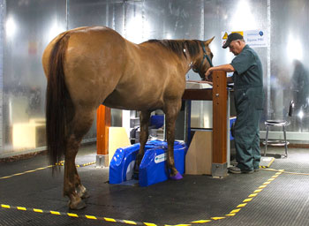
Western Canadian horse owners and referring veterinarians have access to a powerful diagnostic tool: a standing MRI unit in the WCVM Veterinary Medical Centre's Ryan/Dubé Equine Performance Centre. The system allows medical imaging teams to:
- quickly produce high-quality clinical images of the equine foot, pastern, fetlock and carpus
- definitively diagnose conditions in both fore and hind suspensory ligaments
- detect soft tissue lesions in the equine foot or conditions such as navicular syndrome that involve tendons and ligaments
- diagnose certain forms of lameness up to and including the knee (carpus) and hock (tarsus)
Unlike other MRI systems, the WCVM's standing MRI unit doesn't require patients to undergo general anesthesia: a horse is only sedated to ensure minimal movement and good image quality.
Besides being more cost effective, the sedation option allows clinicians to avoid the small but significant risk of anesthetizing an equine patient.
Computed Tomography (CT)
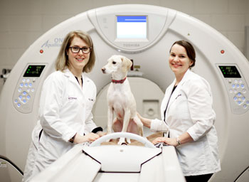
Computed tomography (CT) is a non-invasive imaging method that uses a rotating X-ray beam and computer analysis to produce detailed, cross-sectional images.
One of the technology’s main advantages is its speed: medical imaging specialists can conduct a CT scan on a patient in a matter of minutes.
Medical imaging specialists often use CT technology to:
- quickly assess serious fractures
- identify nasal, ear, sinus or dental diseases
- investigate a patient’s soft tissues or bones for potential tumours, metastasis and other diseases
- identify abdominal conditions
- develop specific radiation therapy plans for veterinary oncology patients
- diagnose and determine treatment options for certain musculoskeletal diseases
- conduct contrast imaging for diagnosing complex disorders
- equine patient specific assessments:
- assess proximal tibia and radius, equine hock and carpus,
- detect injuries of the deep digital flexor tendon or suspensory ligament
- detect potential stress fractures in equine limbs
The CT scanner is part of the WCVM Veterinary Medical Centre's Medical Imaging Department that provides expertise and technology for the centre's Small Animal Clinic and Large Animal Clinic.
CT technology is available to large and small animal patients at the WCVM Veterinary Medical Centre.
Allard-Roozen Imaging Suite
The Allard-Roozen Imaging Centre is home to a positron emission tomography-computed tomography (PET-CT) scanner. The WCVM is one of only five other North American universities with veterinary colleges have a PET-CT unit available for clinical use in animals and for research studies.
A PET-CT scan combines the information available from a CT scan — a three-dimensional X-ray — with a PET scan, which delivers information about the metabolic activity in tissues.
For example, PET-CT technology can help detect cancer, brain disorders, heart disease and infections before any anatomical changes are detectable by other imaging scans.
PET-CT technology relies on the use of radioisotopes. The Allard-Roozen Imaging Suite is located only metres away from the Sylvia Fedoruk Canadian Centre for Nuclear Innovation where the short-lived radioisotopes are produced in the centre’s cyclotron.
The PET-CT technology is available to small animal patients at the WCVM Veterinary Medical Centre.
Nuclear Scintigraphy
Service update, October 15, 2025: the VMC's nuclear scintigraphy equiment is out of service until further notice.
Nuclear scintigraphy is available to large and small animal patients at the WCVM Veterinary Medical Centre.
With nuclear scintigraphy, medical specialists inject a radioactive isotope that's attached or tagged to drugs (or markers). The radioisotopes are absorbed in increased amounts in areas of the body where there's elevated metabolic activity in soft tissue or bone. The specialists then use a gamma camera to measure the level of radioactivity and to produce a two-dimensional image of the patient's body.
Medical imaging specialists use nuclear scintigraphy for large animals to:
- visualize an animal's proximal limbs, back and pelvis
- detect lameness in multiple limbs
- detect suspensory ligament-related injuries
- monitor healing of fractures
- detect and diagnose abnormal growths (benign or malignant)
Medical imaging specialists use nuclear scintigraphy for small animals to:
- diagnose tumours and to search for new growth or spread of tumours
- conduct bone scans in dogs diagnosed with osteosarcoma
- measure kidney function in cats and dogs
- diagnose portosystemic shunts in livers
- evaluate lung perfusion
- conduct lymphatic studies
- conduct companion animal health research for vascular, muscular and skeletal studies
Our Team
The WCVM Veterinary Medical Centre's clinical team members are dedicated, compassionate people with specialized training and a diverse range of experiences. In addition to providing our patients with high-quality care and support, we are helping to train Western Canada's next generation of veterinary professionals.
What to Expect
While the WCVM Veterinary Medical Centre shares many similarities to other veterinary hospitals, there are some differences that may have an impact on your appointment.
Appointments
Animal Owners
Contact us to make an appointment. If you are a new client, please click below to find information about our location, parking and what to expect during your animal's appointment.
Emergency services available 24/7
Emergency services are available for acutely ill or seriously injured animals.