Vet and Owner Info
Information for Referring Veterinarians
If you have referred an animal for cataract surgery, there are a few important things you should know and do to give the animal the best chance at a positive outcome.
Information for Owners
This webpage provides answers to frequently asked questions about cataracts and cataract surgery. This information is also available as a pdf version.
Cataracts

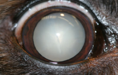
A cataract is any opacity within the lens of the eye. The lens is a disc-shaped structure that is suspended in the eye by tiny ligaments behind the iris (the colored part of the eye). It is normally clear to allow light to enter the eye and it helps to focus that light on the retina. The lens is made up of highly organized cells called lens fibers and is covered by a thin clear capsule that is about the consistency of cellophane.
Cataracts occur when there is damage to the lens fibers. Cataracts may come in many shapes and sizes. They can be small and may not significantly interfere with vision, or they may take up a larger portion of the lens causing obstruction of the light entering the eye and, therefore, blindness.
Cataracts may progress through several different stages. Often, proteins will leak from the lens causing inflammation inside the eye. This can cause discomfort and complications such as glaucoma, retinal detachment and degeneration, all of which may cause permanent blindness.
Causes
In general, cataracts are the result of biochemical damage to the lens fibers which may occur due to several factors:
- Genetics: Most cataracts seen in dogs are inherited. They typically occur in young to middle aged purebred dogs.
- Diabetes: Diabetes mellitus is a common cause of cataracts in dogs. Studies suggest that 68-75% of diabetic dogs will develop cataracts within one year of being diagnosed with diabetes.
- Age: Age related cataracts occasionally occur in animals. They usually do not appear before 6 years in large breed dogs, and 10 years in small breed dogs. These cataracts are often small and progress slowly requiring many months to several years to produce vision loss.
- Miscellaneous causes: Less commonly, cataracts can be caused by toxins, dietary deficiencies, injury of the lens and other ocular diseases such as retinal degeneration, inflammatory conditions and glaucoma.
Treatment
Cataracts can only be removed by surgery. There is no medication that will return the lens back to a clear structure.
Cataract Surgery
Is my dog a candidate for cataract surgery?
In order to determine the answer to that question, your dog will need to have his/her eyes evaluated by a veterinary ophthalmologist. We need to determine that your pet will have a reasonable chance of vision after the surgery has been performed. In addition to an eye examination, some diagnostic tests may be required. Typically an electroretinogram (ERG) is performed to assess the function of the retina and occasionally, an ultrasound of the eye may be required.
Before surgery
If your pet is deemed an appropriate candidate for cataract surgery, a date will be set for the surgery. Because cataracts cause inflammation inside the eye, you will need to treat your pet with a topical antiinflammatory drop at least twice daily after diagnosis of the cataracts.
Please note that your pet should not be bathed or groomed for several weeks after surgery, therefore, plan to have any necessary grooming done before the surgery date.
Skin infections and dental disease can increase the risk of infection after cataract surgery. Skin infections must be resolved prior to surgery. If your pet has dental disease, a dental cleaning should be performed at least 4 weeks prior to the scheduled cataract surgery. It is therefore recommended that you have your pet’s skin and teeth evaluated by your veterinarian before surgery is even considered.
If your dog is diabetic: Diabetic dogs should be well regulated prior to surgery and a urinalysis must be done at your veterinary clinic one week prior to your surgery appointment. This is important as surgery will not be performed if your pet has ketones in the urine or a urinary tract infection and these conditions will require treatment prior to surgery.
Your pet will be examined the day prior to cataract surgery (usually a Monday) and pre-operative bloodwork and an ERG will be performed. If these are within normal limits the surgery will be completed the following day. The morning after surgery your pet will require a re-evaluation. Therefore, you should expect to visit the clinic on 3 consecutive days. Your pet will usually not be hospitalized at night and will be allowed to spend the evenings at home (or in the hotel) with you during this period of time.
The surgical procedure: phacoemulsification
Cataract surgery involves removal of the lens fibers. In order to access the lens, an incision is made in the cornea and a round hole is torn in the front of the lens capsule. A small pen-like instrument is introduced into the eye and through the hole in the lens capsule. This instrument produces ultrasonic vibrations that break up the lens material (phacoemulsification) as it simultaneously vacuums this material to remove it from the eye
After the lens fibers are removed, a lens implant may be inserted into the lens capsule. The lens implant is not necessary for vision, but significantly improves near vision. At the completion of the procedure, the cornea is then closed with tiny stitches that absorb over the next four weeks.
Post-operative care
Following cataract surgery you will have a lot of work to do to help achieve a good outcome for your pet. For the first three weeks after surgery, a protective Elizabethan collar (cone) must be worn at all times to prevent self-mutilation and accidental ocular trauma.
Patients must be severely restricted in their activities and avoid any running, jumping, excess barking or rough play. Several (usually four) different types of eye drops will need to be administered four times daily for the first three weeks and then the frequency of administration will gradually be reduced.
Many dogs will remain on anti-inflammatory eye drops once to twice daily for several months following surgery. Some will require them for life.
Follow-up care
In most cases your pet will be discharged into your care the afternoon of surgery. You will be required to return for a follow-up appointment the next day. Further follow-up times will be decided on an individual basis, but are typically weekly for the first three weeks, then at six, and 12 weeks, six months and 12 months following surgery, then yearly with an ophthalmologist for life.
Success rate
Postoperative success rates for vision following canine cataract surgery are about 80 per cent for the first 2.5 years. However, regardless of the surgical method used to remove the cataract, several postoperative complications can develop. Some of these complications lead to permanent blindness and ocular pain. In those cases further surgery will be required to remove the eye or to place an intrascleral prosthesis.
Possible Complications
- Transient increases in intraocular pressure have been reported to occur in 50 per cent of dogs for 12-72 hours after surgery. The intraocular pressure is monitored after surgery and pressure elevations are treated. Most dogs respond to treatment to reduce the pressure.
- Persistent increases in intraocular pressure (glaucoma) may develop days, weeks, months, or even years after surgery. This type of glaucoma usually forms as a result of permanent structural changes in the region of the eye where fluid normally exits. Glaucoma causes permanent vision loss and discomfort. In some cases it can be controlled with anti-glaucoma medications for a period of time but most cases require removal of the eye or placement of an intrascleral prosthesis. Elevations in intraocular pressure need to be addressed urgently as the longer the pressure is elevated, the more likely the eye is to go blind requiring eye removal.
- Retinal detachments can occur postoperatively. Some can be surgically repaired, however, the prognosis for vision is guarded. The prevalence of retinal detachments has be reported to be low and may be decreased with early cataract removal. Retinal detachment has been reported to be more frequent in certain breeds such as the Bichon Frise, Boston terrier, and Shih Tzu. If untreated, retinal detachments often lead to glaucoma, which often requires removal the eye or placement of an intrascleral prosthesis.
- Opening of the incision can occur secondary to trauma, which can be self-induced and will require surgery to repair.
- Decreased tear production is not necessarily a complication from surgery itself but it may be noted after surgery. Normally, tears cover the corneal surface and are important in lubricating the cornea. The tears are measured by Schirmer tear test strips. When the tear production decreases (“dry-eye”) the cornea is at risk for ulceration and treatment to stimulate tear production is required. This treatment is often necessary for the life of the animal.
- Corneal ulcers can occur following surgery and may be due to exposure and drying of the corneal surface during and after surgery. The healing of such ulcers can be delayed due to decreased tear production, and the administration of anti-inflammatory drops (which are necessary after surgery). Some ulcers can heal with medicine alone but others require surgical repair (a graft) under general anesthesia.
- Uveitis is inflammation inside the eye. This is the most common post-operative complication and is essentially unavoidable after intraocular surgery. Usually, this inflammation will begin to subside within three weeks, however, low grade inflammation can persist for weeks to months. This complication is treated for several weeks after surgery with topical drops and occasionally oral anti-inflammatory tablets.
- Scarring of the lens capsule may be present at the time of surgery or develop after surgery. Scars are more common in mature or hypermature cataracts i.e. those that have been present for a longer period of time.
- Bleeding in the eye can occur during or after surgery due to sudden changes in pressure inside the eye, trauma to the iris during surgery, severe intraocular inflammation, pre-existing bleeding tendencies or retinal tears. Common contributors to bleeding include excessive barking, vigorous activity, head shaking, blunt trauma, tension around the neck (from a collar and leash), and retinal detachment. Thus, minimal activity is essential and excitement must be avoided. A harness instead of a collar is often beneficial for your pet after surgery to avoid excessive tension around the neck.
- Corneal edema (fluid in the cornea) can result from damage to the inner layer of the cornea or from glaucoma. Edema causes the cornea to become cloudy (bluish), which can affect vision. This is more common in older dogs and in certain breed such as the Boston Terrier.
- Infection is essentially a risk of any surgical procedure but is particularly devastating when it occurs in the eye. Infection in the eye usually results in the need to remove the eye.
Appointments and Referrals
Animal Owners
This is a referral-based service. You will need to have your veterinarian submit a referral form to the Veterinary Medical Centre before you can make an appointment with this clinical service.
Your local veterinarian may choose to consult with the WCVM ophthalmology service regarding your animal’s condition and possible treatment protocols. At that point, your veterinarian may decide to refer your animal to the VMC's ophthalmology service.
If you have any questions about a referral patient, please contact WCVM Ophthalmology Service:
- Email: wcvmeyevet@gmail.com
- Tel: 306-966-7126
Emergency services available 24/7
Emergency services are available for acutely ill or seriously injured animals.

