Hard to swallow: USask veterinary team helps pup with feeding issue
When her four-month-old puppy named King began regularly spitting up his food after eating, Angela Seymour knew her little American French bulldog needed help from a veterinarian.
By Jessica Colby“He was really sick, and I took him in,” says Seymour.
The young puppy stayed at a veterinary clinic in Prince Albert, Sask., for several days. But when King continued having swallowing issues after returning home, Seymour and her family decided to take the young dog to the Western College of Veterinary Medicine’s (WCVM) Veterinary Medical Centre (VMC) in Saskatoon, Sask.
Dr. Daniel Moreno (DVM), a resident in small animal internal medicine at the WCVM, took on King’s case at the VMC in early February. Once As a first step, the clinical team performed a swallow study on King to determine what was causing the puppy to spit up — or regurgitate — his food. A swallow study is a type of examination that uses medical imaging to follow the path of food from a pet’s mouth to its stomach.
“After looking at how he behaved after swallowing food, we realized that he was most likely having a problem within his esophagus,” says Moreno. The swallow study confirmed that King had some narrowing in the middle of his esophagus — the tubular organ that runs from the throat to the stomach.
An esophageal stricture is an abnormal narrowing of the esophagus’s inner open space. The condition can affect dogs of all ages and can be caused by several factors — including gastroesophageal reflux.
“Because of the location and [dog’s] age, we had to exclude the possibility of a congenital malformation in his vessels that could be encircling the esophagus, causing a restrictive obstruction there,” says Moreno.
To rule out this problem, the team performed a computed tomography (CT) scan of the puppy. During a CT scan, highly detailed three-dimensional images are taken to aid veterinarians in making a diagnosis.
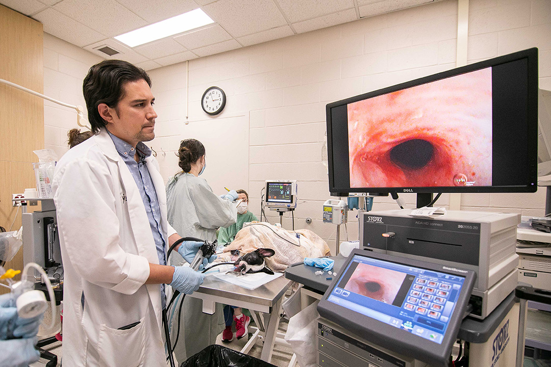
The CT scan showed no vessel malformation, so the veterinary team recommended the next step to Seymour: performing an endoscopy. For this procedure, veterinarians inserted the tube-like instrument into King’s esophagus. The endoscope — equipped with a tiny camera and light — allowed clinicians to get a close-up view of the narrowing in the young dog’s esophagus.
Using a plastic catheter with a deflated balloon on the end, the veterinary team inserted the device into King’s esophagus and gradually inflated the balloon with subsequently increasing amounts of pressure to widen the narrowing.
King returned home with his family the next day, but the puppy did have to return for a second balloon dilation procedure. Now eight months old, King is more active than ever and “he’s doing really good,” according to Seymour, who is very thankful to the entire VMC clinical team for helping her family’s beloved pet.
She says King is recovering well at home and hasn’t experienced any further swallowing issues as he eats his daily rations of wet dog food.
“He’s my baby,” says Seymour.

